Winkelwagen
U heeft geen artikelen in uw winkelwagen
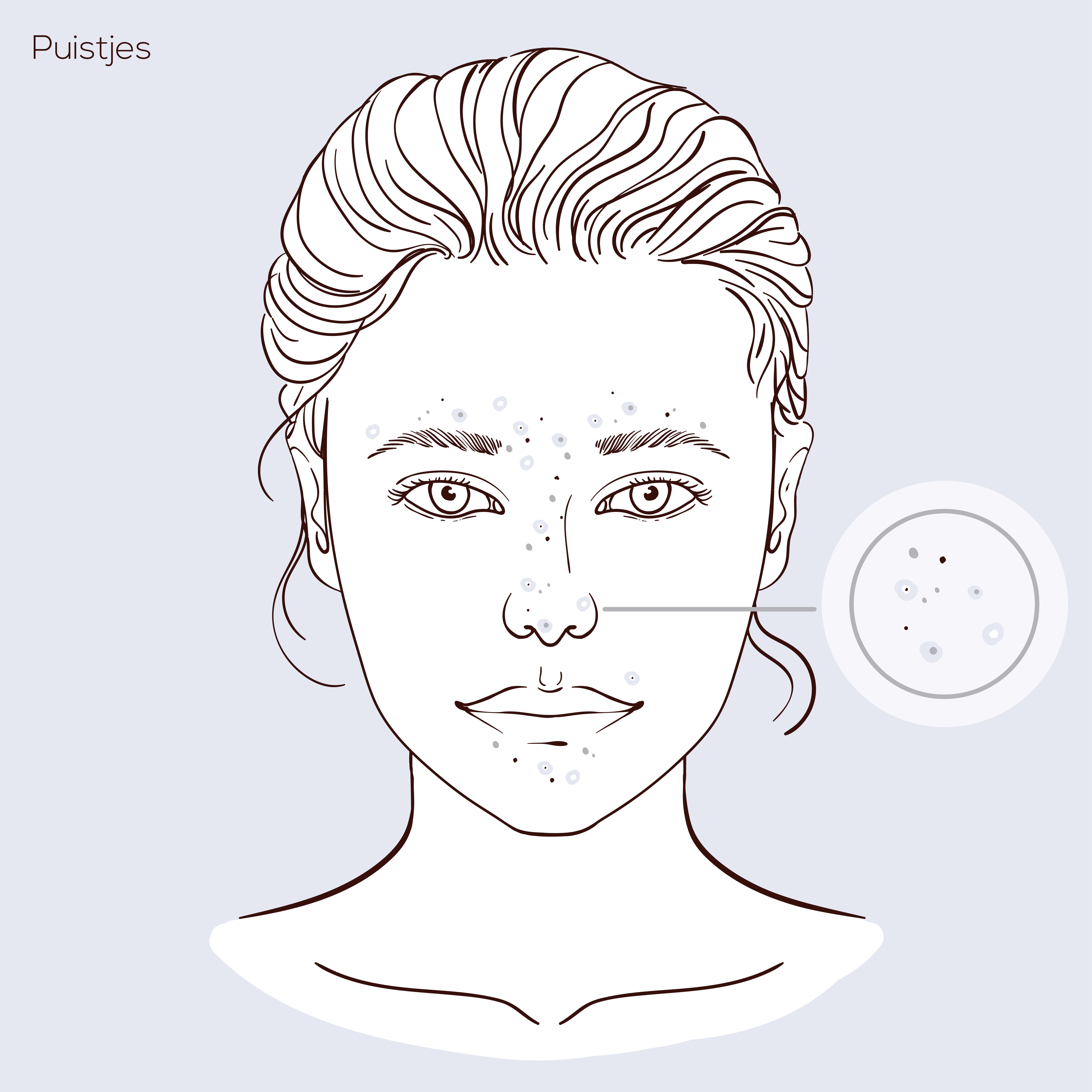
Puistjes komen veel voor. Met name jong volwassenen en tieners hebben er makkelijk last van. Aanleg en hormonen spelen daarin een grote rol. Eén ding is in elk geval zeker: veel mensen ervaren ongemak van puistjes. Iedereen wil immers een mooie, gave huid. In dit artikel bespreken we daarom alle tips, zowel qua leefstijl als cosmetica, om puistjes te voorkomen en te genezen. Mee-eters of puistjes kunnen namelijk allerlei oorzaken hebben en er zijn verschillende manieren om ze tegen te gaan. Hieronder lees je hoe!
We hebben het tot nu toe vooral over puistjes in je gezicht gehad. Toch komen ze bij veel mensen ook op de rest van het lichaam voor, zoals op de rug, schouders en soms zelfs op de billen en bovenbenen.
Puistjes op je lichaam zijn in die zin minder vervelend, dat je ze wat makkelijker kan verbergen. Wel zijn ze vaak hardnekkiger. Dat heeft met name te maken met het dragen van je kleding, want dat zorgt voor warmte en wrijving op de huid. Puistjes blijven daardoor makkelijker geïrriteerd en verdwijnen minder snel.
Wat kan je doen tegen puistjes op je lichaam:
Puistjes en mee-eters komen bij veel mensen voor op het voorhoofd. Bij veel mensen is de huid daar wat vetter. Puistjes ontstaan er daardoor sneller. Je kan hiervoor gewoon de tips gebruiken die we al eerder in deze blog genoemd hebben.
Bij een aantal mensen zijn puistjes op het voorhoofd niet de standaard puistjes of mee-eters, maar gistpuistjes. Gistpuistjes zijn vaak niet erg opvallend. Bij veel mensen zijn ze niet rood, geïrriteerd of pijnlijk. Wel kunnen ze soms jeuken. De één heeft meer aanleg voor gistpuistjes op het voorhoofd dan de ander.
Wat kan je doen tegen (gist) puistjes op je voorhoofd:
Puistjes op je hoofd
Puistjes bovenop je hoofd zijn vaak niet hetzelfde als de acne puistjes die het meest voorkomen. Op je hoofd kunnen puistjes lastig te bestrijden zijn, vanwege je haar.
Puistjes op het hoofd hebben vrijwel altijd een bacterie of schimmel als oorzaak. Het is aan te raden om hiervoor naar de huisarts te gaan, omdat er mogelijk anti-schimmel of antibacteriële medicatie nodig is.
Puistjes op je hoofd kunnen ook veroorzaakt worden door overgevoeligheid voor cosmetica. Ben je een nieuw product gaan gebruiken en is het probleem daarna ontstaan, dan zou je kunnen kijken wat er gebeurt als je het desbetreffende product weglaat.

Puistjes, mee-eters en acne waren tien jaar geleden nog vrij lastig te behandelen. Inmiddels zijn er echter uiteenlopende effectieve medicijnen en behandelingen op de markt om acne, puistjes en mee-eters efficiënte te verhelpen.
Zo zijn er tegenwoordig antibiotica verkrijgbaar die de Propionibacterium Acnes (P.acne) bacterie en Staphylococcus Aureus bacterie doden. Dit zijn de bacteriën die verantwoordelijk zijn voor acne-gerelateerde huidinfecties. Bovendien zijn er inmiddels hormoonremmers op de markt die je hormoonhuishouding stabiliseren. De belangrijkste voorbeelden van effectieve medicijnen tegen acne, puistjes en mee-eters zijn als volgt:
Benzoylperoxide is een zeer effectieve en werkzame stof ter behandeling van puistjes en acne. Benzoylperoxide wordt verwerkt in gel en crème tegen puistjes zoals Benzac® en Akneroxid®. Benzoylperoxide weekt het opperlaagje van de huid los en remt de groei ervan. Bovendien zorgt benzoylperoxide ervoor dat de ‘Propionibacterium acnes’ bacterie (P. acnes) en Staphylococcus Aureus bacterie zich niet kunnen vermenigvuldigen.
Antibiotica gaan bacteriële infecties tegen en remmen de vermenigvuldiging van onder meer acne-gerelateerde bacteriën. Zodoende kan een antibioticum een effectief medicijn vormen tegen puistjes en mee-eters. De bekendste antibiotica tegen huidklachten, gelieerd aan acne zijn als volgt:
Vitamine A-zuur kan een enorm positief effect hebben op je huid. Daarom worden er in allerlei crèmes, zalven en lotions voor de huid verscheidene derivaten van vitamine A gebruikt. De bekendste hiervan zijn:

Heb je de huid goed gereinigd met een mild product op basis van jouw huidtype? Dan is het belangrijk dat je de huid na de reiniging ook goed verzorgd. Na het sluiten van de poriën is het belangrijk om de huid te hydrateren.
Doe dit bijvoorbeeld met een serum en een crème. Het serum is een effectief product die uit pure werkstoffen bestaat. Nadat de huid gereinigd is zal het serum direct door de huid op worden genomen. Het serum zal diep in de huid kunnen dringen, waardoor het goed zijn werking kan doen. Door het serum af te stemmen op jouw huidtype, zal het ervoor zorgen dat de huid goed verzorgd wordt op de juiste manier.
Een serum is een dun product, waardoor de huid het product direct op kan nemen. Je zal na het aanbrengen van het serum dan ook geen zacht gevoel krijgen. Daarom is het belangrijk om na het aanbrengen van het serum een crème te smeren. Een crème zorgt voor comfort en ook zal deze een effectieve werking hebben voor de huid. Na het aanbrengen van de crème zal de huid fijn, zacht en soepel aanvoelen. Bepaal ook weer op basis van jouw huidtype welke crème het beste gebruikt kan worden na jouw gezichtsreiniging.
Na het uitvoeren van jouw gezichtsreiniging is het belangrijk om de huid te hydrateren. Door de huid te hydrateren zal je ervoor zorgen dat jouw huid het vocht beter en langer vast kan houden. Zo zullen de cellen in de huid minder snel het vocht kwijtraken, waardoor de huid minder snel droog en trekkerig aanvoelt. Hydrateer de huid altijd goed, zodat het vochtgehalte op peil kan worden gehouden en de huid niet uit balans draait.

Zoals gezegd is reinigen de basis van een goede skincare routine, maar waarom is deze stap zo belangrijk? We hebben de vier belangrijkste redenen voor je op een rijtje gezet:
1. Om het alledaagse vuil te verwijderen
Het laagje vuil dat zich op je huid bevindt, verdwijnt niet door je gezicht alleen te spoelen met water. Je huid heeft meer reiniging nodig dan dat. Daarnaast droogt water je huid uit en kan je beter een reiniger gebruiken die ook verzorgend is.
2. Om onzuiverheden te voorkomen
Door te reinigen voorkom je verstopte poriën en een doffe huid. Vooral bij mensen met een vet huidtype kunnen mee-eters en puistjes ontstaan als het gezicht niet goed gereinigd wordt. Bij een hoge talgproductie hebben poriën de neiging sneller verstopt te raken of kunnen ze grof zijn. Een goede reiniging voorkomt deze onzuiverheden.
3. Om actieve ingrediënten beter in te laten trekken
Na een goede reinigingsbeurt gebruik je waarschijnlijk nog andere gezichtsverzorgingsproducten, zoals een serum of een crème. De actieve ingrediënten in deze producten moeten goed in je huid kunnen trekken voor een optimaal resultaat. Dit kan niet wanneer je huid verstopt zit onder een laagje vuil of wanneer je poriën verstopt zijn.
4. Om een doffe huid te voorkomen
Een doffe huid kan ook het gevolg zijn van een slechte reiniging of reiniging met te agressieve producten. Een goede reiniger zorgt ervoor dat je dode huidcellen loslaten, waardoor de jonge huidcellen zich sneller vernieuwen. Dit zorgt voor meer glans in je gezicht.
