Winkelwagen
U heeft geen artikelen in uw winkelwagen
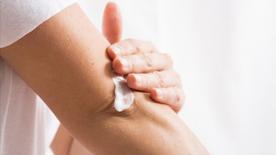
Eine Bodylotion ist eine Emulsion aus Wasser und Fett, die die Haut nach dem Duschen oder Baden pflegt und über mehrere Stunden mit Feuchtigkeit versorgt. Es gibt ein großes Angebot unterschiedlicher Körperlotionen auf dem Markt, die wir in unserem Bodylotion-Test genauer betrachten. Beachten Sie, dass verschiedene Hauttypen unterschiedliche Bedürfnisse haben und wählen Sie Ihre Bodylotion Ihrem Hauttyp entsprechend aus.
Welche Bodylotion ist die beste?
Die Frage, welche Bodylotion die beste ist, lässt sich pauschal nicht beantworten, denn trockene oder sensible Haut benötigt beispielsweise eine andere Pflege als normale Haut. Welche Creme die beste für Sie ist, hängt also auch von Ihrem Hauttyp ab. Dennoch gibt es allgemeine Kriterien, an denen Sie eine gute Bodylotion erkennen. So sollte die Creme pflegende und feuchtigkeitsspendende Inhaltsstoffe enthalten und auf toxische und potenziell hautreizende Inhaltsstoffe verzichten. Welche das sind und welche Körperlotionen empfehlenswert sind, haben wir in unserem Bodylotion-Test für Sie aufgelistet.
Per Dr. Idriss's earlier point about addressing specific skin conditions, you can also take those into account. For example, she says formulas that contain soothing colloidal oatmeal are an especially good choice for those battling itchiness, irritation, or even eczema. "I also recommend lotions with niacinamide for those who have back acne, since it not only helps maintain the lipid barrier of the skin but is also anti-inflammatory," she adds.
A word on ingredients. The three most common categories found in moisturizers, of any kind, are humectants, emollients, and occlusives, each of which functions in a slightly different way.
Ideally, you do want a combination of all three in a good moisturizer formulation, though again, exactly how much of each depends on exactly how dry your skin is.

Je höher der Fettanteil der Emulsion, desto reichhaltiger ist die Creme. In der Produktbezeichnung steckt in manchen ein Hinweis auf das Mischungsverhältnis von Wasser und Fett. Generell haben Köperlotionen einen höheren Wasseranteil, deshalb sind sie eher für normale Haut geeignet. Pflegeprodukte für trockene Haut werden gerne als Bodymilk, Körpercreme und Balsam bezeichnet, sie haben einen höheren Fettanteil.
Bodybutter wurde ursprünglich aus reinen Wachsen und Fetten gemacht. Sie war bei Raumtemperatur fest und ließ sich erst durch das Erwärmen in den Händen verflüssigen. So konnte die Butter dann auf die Haut aufgetragen und einmassiert werden. Mittlerweile besteht die Körperbutter meist aus sehr gehaltvollen Emulsionen. Ihr Hauptbestandteil sind heute flüssige Öle. Sie enthalten aber auch Wasser und müssen daher – im Gegensatz zu traditionellen Körperbutterstücken – konserviert werden.

Ökotest hat insgesamt 35 Körperlotionen für trockene Haut miteinander verglichen. Im ersten Schritt prüften die Tester die Deklaration auf der Verpackung: Sind bedenkliche Inhaltsstoffe wie beispielsweise BHT oder PEG/PEG-Derivate vorhanden?
Im zweiten Schritt wurden die Lotionen in spezialisierten Laboren auf synthetische Schadstoffe wie Formaldehyd überprüft und auf natürliche Duftstoffe wie Cinnamal, die Allergien verursachen können. Bei Verdachtsfällen wurde zusätzlich der Gehalt auf MOAH und Parabenen gemessen.
Schließlich interessierten sich die Tester für die Verpackung: Ist sie bei Glasbehältern bruchsicher verpackt? In welchem Anteil wurde Recyclingmaterial für die Plastikverpackung verwendet und wurde das korrekt deklariert?

Jean & Len Reichhaltige Bodymilk
Die Reichhaltige Body Milk mit Sheabutter und Vitamin E des deutschen Newcomer-Labels Jean & Len pflegt trockene Haut intensiv und ist dabei besonders hautverträglich. Die Lotion enthält keine Parabene, Silikone, Mikroplastik oder Mineralöl und ist außerdem vegan. Beim Verbrauchermagazin ÖKO-Test schnitt die Körperlotion deshalb auch mit „sehr gut“ ab. Die Lotion zieht schnell ein und sorgt für ein langanhaltend geschmeidiges Hautgefühl. Extra-Pluspunkt: Der sanfte Duft der Sheabutter verwöhnt die Sinne beim Eincremen.
Jean & Len Reichhaltige Body Milk im Überblick:
Das sagen Nutzer:innen: Bei Amazon erreicht die Body Milk sehr gute Bewertungen. Laut Nutzer:innen zieht die Bodylotion schnell ein und sorgt für ein langanhaltend gutes und gepflegtes Hautgefühl – auch bei Tester:innen mit sehr trockener Haut. Auch der angenehme Duft wird oft gelobt.
Sheabutter ist das Fett aus den Sheanüssen des Sheabaums. Sie wirkt rückfettend, feuchtigkeitsbindend und entzündungshemmend und ist auch für die Pflege sehr trockener oder gereizter Haut geeignet.
