
Winkelwagen
U heeft geen artikelen in uw winkelwagen
If present, the underlying systemic disease must be treated, but there is not an accepted correlation between the underlying systemic disease severity and the severity of pyoderma gangrenosum.[11][2]
The use of systemic immunosuppression is determined by how fast the disease progresses. If the size of the lesion is rapidly growing, systemic medications like corticosteroids or cyclosporine may be used. The STOP GAP randomized control trial in 2015 compared oral prednisolone and oral cyclosporine in the treatment of pyoderma gangrenosum. The results did not show a significant difference in the speed of lesion healing with either medication. At six months, 47% of patients on cyclosporine had healed ulcers, and 47% of the patients on prednisolone had healed ulcers. Recurrence was similar among the two groups as were adverse reactions. However, serious adverse reactions such as infections were more common in the patients receiving prednisolone.
For more indolent or limited disease, topical or intralesional therapy alone may be sufficient. Topical and intralesional steroids, as well as topical tacrolimus, have been used with success. Other agents that have been tried are nicotine, topical dapsone, and sodium cromoglycate.
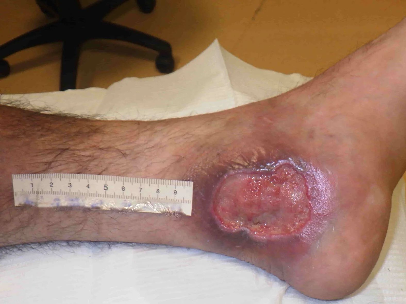
Wound care and pain control are key features in the treatment of pyoderma gangrenosum. Patients and healthcare providers must cleanse the wound to prevent infection. Debridement is important but must be performed very cautiously to ensure that only nonviable tissue is removed because of the aforementioned association with pathergy.
Other therapies that have been successful are anti-TNF-alpha drugs such as etanercept and adalimumab. Ustekinumab, an IL-12/23 inhibitor used in the treatment of psoriasis, has been reported to improve pyoderma gangrenosum. Other therapies that may be effective in the treatment of pyoderma gangrenosum include Canakinumab, an IL-1 beta monoclonal antibody, and tocilizumab, an anti IL-6 monoclonal antibody.
1 Department of Cosmetology and Esthetic Dermatology, Ludwik Rydygier Medical College in Bydgoszcz, Nicolaus Copernicus University in Torun, Poland. Head of the Department: Prof. Barbara Zegarska MD, PhD
2 Department of Dermatology, Sexually Transmitted Diseases and Immunodermatology, Ludwik Rydygier Medical College in Bydgoszcz, Nicolaus Copernicus University in Torun, Poland. Head of the Department: Rafał Czajkowski MD, PhD, DSc
1 Department of Cosmetology and Esthetic Dermatology, Ludwik Rydygier Medical College in Bydgoszcz, Nicolaus Copernicus University in Torun, Poland. Head of the Department: Prof. Barbara Zegarska MD, PhD
3 Department of Dermatology and Pathology, Rutgers University New Jersey Medical School, Newark, New Jersey, USA. Head of the Department: Prof. Robert A. Schwartz
4 Department of Radiology and Diagnostic Imaging, Ludwik Rydygier Medical College in Bydgoszcz, Nicolaus Copernicus University in Torun, Poland. Head of the Department: Zbigniew Serafin MD, PhD, DSc
5 Laboratory of Cell Biology and Genetics, Ludwik Rydygier Medical College in Bydgoszcz, Nicolaus Copernicus University in Torun, Poland. Head of the Department: Dorota Olszewska-Słonina MD, PhD, DSc
2 Department of Dermatology, Sexually Transmitted Diseases and Immunodermatology, Ludwik Rydygier Medical College in Bydgoszcz, Nicolaus Copernicus University in Torun, Poland. Head of the Department: Rafał Czajkowski MD, PhD, DSc
1 Department of Cosmetology and Esthetic Dermatology, Ludwik Rydygier Medical College in Bydgoszcz, Nicolaus Copernicus University in Torun, Poland. Head of the Department: Prof. Barbara Zegarska MD, PhD
2 Department of Dermatology, Sexually Transmitted Diseases and Immunodermatology, Ludwik Rydygier Medical College in Bydgoszcz, Nicolaus Copernicus University in Torun, Poland. Head of the Department: Rafał Czajkowski MD, PhD, DSc
The authors declare no conflict of interest.
1. Birnkrant MJ, Papadopoulos AJ, Schwartz RA, et al. Pyoderma gangrenosum, acne conglobata, and IgA gammopathy. Int J Dermatol. 2003, 42 :213–6. [PubMed] [Google Scholar]
2. Jablonska S. Zur Pathogenese des pyoderma gangrenosum. Hautarzt. 1964, 15 :584–91. [PubMed] [Google Scholar]
3. Jablonska S, Stachow A, Dabrowska H. Rapports entre la pyodermite gangreneuse et le myelome. Ann Dermatol Syphilol (Paris) 1967, 94 :121–32. [PubMed] [Google Scholar]
4. Su WP, Davis MD, Weenig RH, et al. Pyoderma gangrenosum: clinicopathologic correlation and proposed diagnostic criteria. Int J Dermatol. 2004, 43 :790–800. [PubMed] [Google Scholar]
5. Bergler-Czop B, Brzezińska-Wcisło L. Pyoderma gangrenosum in a patient with common variable primary immunodeficiency. Postep Derm Alergol. 2013, 30 :188–91. [PMC free article] [PubMed] [Google Scholar]
6. Ruocco E, Sangiuliano S, Gravina AG, et al. Pyoderma gangrenosum: an updated review. JEADV. 2009, 23 :1008–17. [PubMed] [Google Scholar]
7. Binus AM, Quershi AA, Li VW. Pyoderma gangrenosum: a retrospective review of patients characteristics, comorbidities and therapy in 103 patients. Br J Dermatol. 2011, 165 :1244–50. [PubMed] [Google Scholar]
8. Wolska K, Michalska-Jakubus M, Pucuła J, et al. Bullous pyoderma gangrenosum associated with pancytopenia of unknown origin. Postep Derm Alergol. 2014, 31 :272–6. [PMC free article] [PubMed] [Google Scholar]
9. Conrad C, Trued RM. Pyoderma gangrenosum. J Dtsch Dermatol Ges. 2005, 3 :334–42. [PubMed] [Google Scholar]
10. Langan SM, Powell FC. Vegetative pyoderma gangrenosum: a report of two new cases and a review of the literature. Int J Dermatol. 2005, 44 :623–9. [PubMed] [Google Scholar]
11. Braun-Falco M, Kovnerystyy O, Lohse P, et al. Pyoderma gangrenosum, acne and suppurative hidradenitis (PASH) – a new autoinflammatory syndrome distict from PAPA syndrome. J Am Acad Dermatol. 2011, 66 :409–15. [PubMed] [Google Scholar]
This is an Open Access article distributed under the terms of the Creative Commons Attribution-NonCommercial License (http://creativecommons.org/licenses/by-nc/4.0/), which permits unrestricted non-commercial use, distribution, and reproduction in any medium, provided the original work is properly cited.
A 71-year-old-male with hypertension, type 2 diabetes mellitus, morbid obesity, and diastolic heart failure presented to the hospital with mild symptoms of sore throat, fevers, and shortness of breath and tested positive for COVID-19 by RT-PCR. He did not require admission and was discharged home with instructions to self-quarantine for 14 days. About 10 days after diagnosis, he developed painful and pruritic pustules on his left scrotum that quickly ulcerated within a few days. After self-treatment with over-the-counter antifungal and antibiotic creams, the patient was treated at an urgent care facility with cephalexin 500 mg three times daily and valacyclovir 1000 mg twice daily due to concern for cellulitis and shingles. Over the next few weeks, the small ulcers coalesced to form a large painful ulcer ( Figure 1(a )), followed by the appearance of a similar lesion in the lower abdomen ( Figure 1(b )).
Ulcerated lesions on the scrotum (a) and lower abdomen (b) of the patient
He was prescribed topical triamcinolone by his physician and referred to a wound care center where he was diagnosed with pressure ulcers and treated with topical collagenase, a debriding agent, which worsened the ulcers. The patient gradually developed more ulcers on the penis, groin, buttocks, and abdomen over a span of two to three months. He eventually underwent a lesional biopsy revealing neutrophilic dermatosis with perivascular and interstital neutrophilic infiltrates in the dermis and was diagnosed with pyoderma gangrenosum (PG) based on the clinical presentation and histopathology. The patient was started on 60 mg prednisone daily and topical corticosteroids with prompt improvement of ulcers and he is currently being transitioned to infliximab for long-term treatment. Work up for PG-associated disorders including rheumatoid arthritis and other major autoimmune diseases, MGUS, inflammatory bowel disease, and hematological malignancies were all negative.
A little more than a year after being termed a pandemic, our knowledge about COVID-19 symptoms, presentation, pathogenesis, and treatment continues to evolve rapidly. Several cutaneous manifestations of COVID-19 have been described but the major ones include chilblain-like or pseudo-chilblain lesions, urticarial lesions, morbilliform lesions, varicella-like vesicular lesions, purpuric lesions, and livedoid lesions [1]. The mechanisms behind cutaneous manifestations in COVID-19 are still under investigation, but likely involve the indirect effects of immune system hyperactivity and hypercoagulability.
Several immunological similarities exist between COVID-19 and PG. Proinflammatory cytokines and neutrophilic abnormalities play a significant role in the pathogenesis of both diseases. A high neutrophil-to-lymphocyte ratio is an independent risk factor for severe COVID-19 and neutrophilia is an indicator of poor outcomes in COVID-19 patients [2]. PG’s histological finding of neutrophilic abscesses and suppurative inflammation in the dermis is indicative of neutrophilic dysfunction [3].
Biologics targeting the proinflammatory cytokines TNF-α, Interleukin-12 (IL-12), and IL-23 have been very effective in treating PG [4]. IL-6 plays a key role in the activation and accumulation of neutrophils and potentially plays a significant role in hyperinflammation in both COVID-19 and PG. The use of IL-6 antagonist, tocilizumab, along with systemic corticosteroids is currently recommended by the National Institutes of Health (NIH) COVID-19 treatment guidelines panel for severe or rapidly deteriorating patients [5] and was shown to be effective for PG treatment in one case study [6].