
Winkelwagen
U heeft geen artikelen in uw winkelwagen
Spider angiomas are common skin lesions that get their name due to their spiderlike appearance. Most of the time, they are harmless and will go away on their own. However, if you have multiple spider angiomas and are experiencing other symptoms, such as yellowing of your skin (jaundice) and fluid retention, they can indicate liver disease.
Other contributing factors, such as liver cirrhosis or autoimmune disease, warrant medical diagnosis and treatment of the underlying condition. When in doubt, make an appointment with your medical team to be evaluated.
Verywell Health uses only high-quality sources, including peer-reviewed studies, to support the facts within our articles. Read our editorial process to learn more about how we fact-check and keep our content accurate, reliable, and trustworthy.
By Barbie Cervoni, RD
Cervoni is a New York-based registered dietitian and certified diabetes care and education specialist.
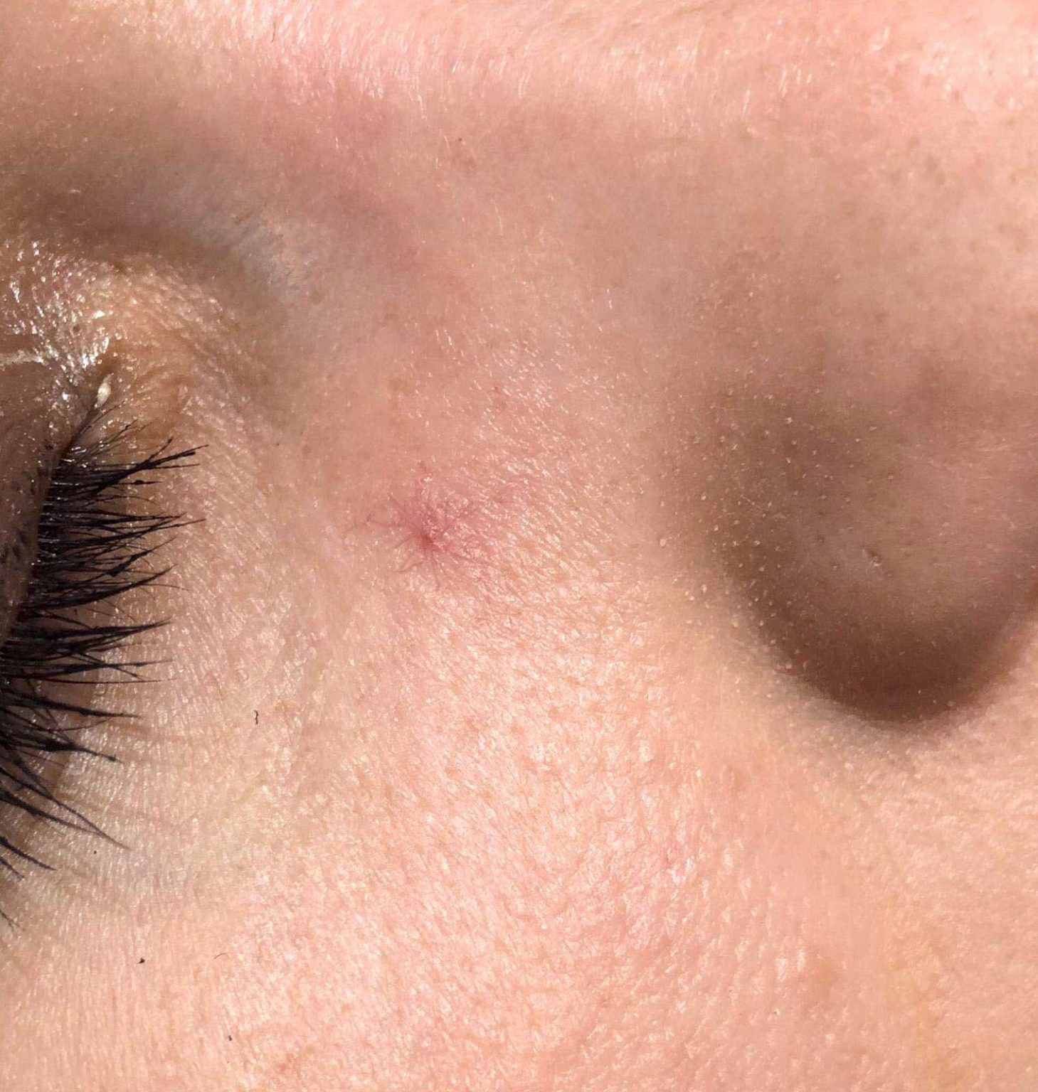
Nevus araneus, also known as spider angioma or spider nevus, is a common benign vascular lesion present in 10-15% of healthy adults and young children. [1, 2] They may appear as a solitary or multiple lesions. [2] In particular, when multiple lesions are present, liver disease, [3] arteriovenous malformations (AVM), [4] estrogen therapy, esophageal and gastric varices, [3] and thyrotoxicosis should be considered. The name stems from its physical appearance, which is characterized by a central red arteriole, or punctum, representing the body of the spider, surrounded by a radial pattern of thin-walled capillaries, resembling legs (see the image below).
--> A spider nevus consists of a central arteriole with radiating thin-walled vessels. Compression of the central vessel produces blanching and temporarily obliterates the lesion. When released, the threadlike vessels quickly refill with blood from the central arteriole. The ascending central arteriole resembles a spider's body, and the radiating fine vessels resemble multiple spider legs.
Nevus araneus lesions range in size from 1-10 mm in diameter. Compression of the central vessel with a slide (diascopy) results in blanching and temporary obliteration of the lesion, which is followed by rapid return of blood flow upon release. [1] Pulsations may occasionally be felt upon compression of the punctum. [5] In adults, these lesions are most frequently found on exposed areas of the body, such as the face, neck, upper trunk (above the nipple line), and arms. In children, the backs of the hands and fingers are commonly affected. [1, 5]

In children, treatment usually is not necessary, and while some lesions resolve spontaneously, others may be permanent. [1] Spider angiomas (nevi araneus) that regress do so over the course of several years.
In young women, lesions often resolve spontaneously within 6 weeks to 9 months after the birth of a child or after discontinuing oral contraceptives. [6]
Numerous lesions associated with liver disease may improve upon treatment of the underlying condition. Reports have described regression after liver transplantation. [11]
Electrodesiccation and laser treatment both can be effective for bothersome facial spider angiomas. Although the risk of a small scar may be slightly higher with electrodesiccation, good results generally are achieved with either intervention. Although cherry angiomas are different vascular lesions, a rater-blinded randomized controlled study of treatment of those showed very little difference between the results of electrodesiccation versus the pulsed dye or KTP laser. [12] Usually, disappearance of the spider angioma follows electrodesiccation. Recurrences are common.
Local anesthesia prior to therapy is optional in adults but advisable in children. Intradermal injection of 0.1-0.2 mL physiological saline solution produces brief complete anesthesia of the site and does not sting on injection. This represents a viable alternative to lidocaine. The central vascular papule has very few nerve endings. Rather than intradermal anesthesia injection, a 30-gauge needle can be inserted directly into the central papule. Anesthesia is flushed into the spider angioma, producing less pain.
Although very common—affecting nearly one in every ten adults—spider angiomas are not considered hereditary and do not run in families. Spider angiomas are very commonly seen in children as well.
For most people, the appearance of spider angiomas are the only presenting symptom and are of no concern. They generally look like a red dot in the center with thin vessels branching out, but may appear differently in your specific case. The thin vessels form a web-like shape and may be red, blue, or even purple in color. Applying pressure to the area will usually cause them to temporarily disappear, reappearing once the blood starts flowing back.
Spider angiomas can appear anywhere on the body, with the face, neck, or sun exposed legs being the most common locations. In rare instances, some people may feel aching or burning around the angioma on the leg that may be associated with long periods of standing.