
Winkelwagen
U heeft geen artikelen in uw winkelwagen
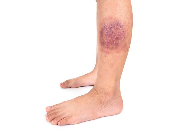
Schimmels en gisten zijn overal. Ze zitten bij iedereen op het lichaam. Dit is meestal niet erg. Op uw huid zit een vetlaagje dat u beschermt en zorgt dat u geen klachten krijgt.
Soms werkt de bescherming van uw huid minder goed. Bijvoorbeeld als het vetlaagje van uw huid eraf gaat. De schimmel komt dan uw huid binnen en kan daar groeien. U krijgt dan plekken op de huid.
In deze situaties krijgt u sneller last van schimmels:
De schimmel komt meestal van iemand in uw omgeving.
De schimmel zit in schilfers van de huid. De schilfers laten los van de huid en komen op bijvoorbeeld de vloeren van zwembaden, doucheruimtes en sportruimtes. Als u deze schilfers op uw huid krijgt, kunt u plekken krijgen.
Sommige schimmels kunt u krijgen van dieren.
In the United States, tinea capitis most commonly affects children of African heritage between three and nine years of age. 4 There are three types of tinea capitis: gray patch, black dot, and favus. Black dot, caused by Trichophyton tonsurans, is most common in the United States (Figure 4). Early disease can be limited to itching and scaling, but the more classic presentation involves one or more scaly patches of alopecia with hairs broken at the skin line (black dots) and crusting. Tinea capitis may progress to kerion, which is characterized by boggy tender plaques and pustules. The child with tinea capitis will generally have cervical and suboccipital lymphadenopathy, and the physician may need to broaden the differential diagnosis if lymphadenopathy is absent. 7 However, lymphadenopathy can also occur in nonfungal scalp disease, and the absence of lymphadenopathy in an otherwise typical presentation should not delay aggressive treatment for tinea capitis. 9
Many physicians treat tinea capitis without a confirmatory culture or KOH preparation if the presentation is typical (i.e., urban setting and child presents with scaling, alopecia, and adenopathy). 2 , 7 , 8 The most common mimics include seborrheic dermatitis and alopecia areata (Table 2). 2 , 3 In atypical cases, a KOH preparation can be performed by scraping the black dots (broken hairs) and looking for fungal spores. The spores of T. tonsurans will be contained within the hair shaft, but for the less common Microsporum canis, the spores will coat the outside of the hair shaft.
A culture, which is more sensitive than the KOH preparation, 10 , 11 can be performed by moistening a cotton applicator or toothbrush with tap water and rubbing it over the involved scalp. The sample is then applied to Sabouraud liquid medium or Dermatophyte test medium. Children with kerion have a high false-negative culture rate. 10 A Wood lamp examination of scalp lesions is often not helpful because the most common cause, T. tonsurans, does not fluoresce. M. canis, which is more common in white children, exhibits a green fluorescence under a Wood lamp. Microsporum infections result from exposure to infected dogs or cats and may produce much more inflammation than Trichophyton infections. 4
JOHN W. ELY, MD, MSPH, SANDRA ROSENFELD, MD, AND MARY SEABURY STONE, MD
Am Fam Physician. 2014,90(10):702-711
Author disclosure: No relevant financial affiliations.
Tinea infections are caused by dermatophytes and are classified by the involved site. The most common infections in prepubertal children are tinea corporis and tinea capitis, whereas adolescents and adults are more likely to develop tinea cruris, tinea pedis, and tinea unguium (onychomycosis). The clinical diagnosis can be unreliable because tinea infections have many mimics, which can manifest identical lesions. For example, tinea corporis can be confused with eczema, tinea capitis can be confused with alopecia areata, and onychomycosis can be confused with dystrophic toe-nails from repeated low-level trauma. Physicians should confirm suspected onychomycosis and tinea capitis with a potassium hydroxide preparation or culture. Tinea corporis, tinea cruris, and tinea pedis generally respond to inexpensive topical agents such as terbinafine cream or butenafine cream, but oral antifungal agents may be indicated for extensive disease, failed topical treatment, immunocompromised patients, or severe moccasin-type tinea pedis. Oral terbinafine is first-line therapy for tinea capitis and onychomycosis because of its tolerability, high cure rate, and low cost. However, kerion should be treated with griseofulvin unless Trichophyton has been documented as the pathogen. Failure to treat kerion promptly can lead to scarring and permanent hair loss.
| Clinical recommendation | Evidence rating | References |
|---|---|---|
| Tinea corporis, tinea cruris, and tinea pedis can often be diagnosed based on appearance, but a potassium hydroxide preparation or culture should be performed when the appearance is atypical. | C | 2 |
| Acceptable treatments for tinea capitis, with shorter treatment courses than griseofulvin, include terbinafine (Lamisil) and fluconazole (Diflucan). | A | 14 – 16 |
| The diagnosis of onychomycosis should generally be confirmed with a test such as potassium hydroxide preparation, culture, or periodic acid–Schiff stain before initiating treatment. | C | 27 |
