Winkelwagen
U heeft geen artikelen in uw winkelwagen
A joint power of attorney is just a legal document that authorizes an agent to act on your behalf when making financial or legal decisions that you normally have to make for yourself.
For example, a bank will not allow someone who is not named on your bank account to withdraw funds from your account. Normally, only you (or another account holder) may access or withdraw funds in your account. If you are not able to do this, by presenting a joint power of attorney that you have signed, family members or friends may be able to perform this task for you.
The joint power of attorney serves as a legal permission slip–it authorizes third-party service providers (like banks) to conduct financial or legal business with the agent as if they were conducting your business with you.
MJ
Attorney, distinguished law professor
Cake values integrity and transparency. We follow a strict editorial process to provide you with the best content possible. We also may earn commission from purchases made through affiliate links. As an Amazon Associate, we earn from qualifying purchases. Learn more in our affiliate disclosure.
Part of establishing an effective estate plan for yourself may be to employ the knowledge, skills, and expertise of others to assist with important decision making.
For example, in attempting to buy or sell a piece of real property, you may want the knowledge of a local real estate agent to help you choose the right property. When considering market investments, you may want to rely on the insights of experienced investment brokers to choose the most prudent investments.

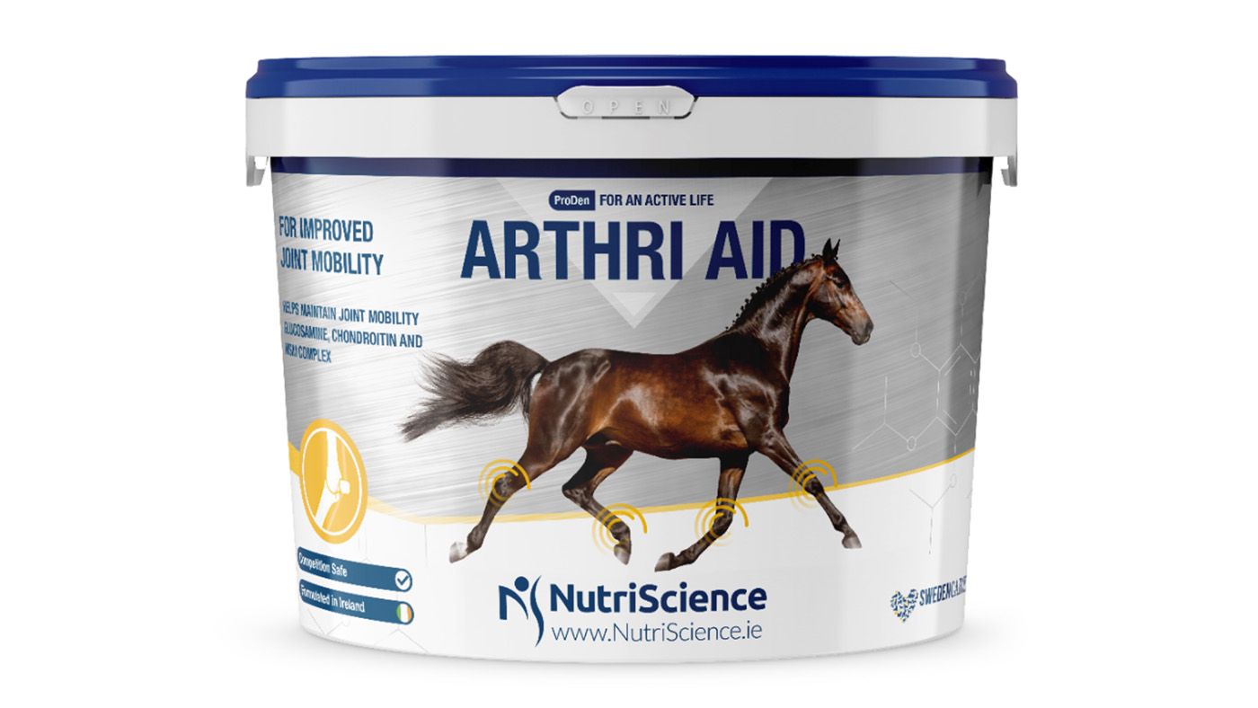
Look out for the most common ingredients found in joint supplements, which includes:
Consider the form the supplement takes – joint supplements can come in liquid, powder and pellet forms. Your horse may have a preference to one over another, which could make all the difference as if he won’t eat it, your horse won’t receive the benefit.
Cost is another consideration – we’ve calculated the daily cost of all the supplements featured in this guide to help you compare them easily. To make it as comparable as possible, we’ve used the smallest tubs available, but if you’re better off buying a larger volume of supplement if you can as the daily cost can become significantly lower.
If you compete with your horse then it is important you make sure any supplements you use do not contain any prohibited substances.
A principle also can name when a joint (or joint and several) power of attorney is effective. A joint power of attorney can be:
There are different situations when you’d use each of these types.
So, if you suffer a mental illness and cannot make financial decisions, your agents will be able to continue managing your financial affairs during your incapacity. A principal may rescind or terminate a durable power of attorney at any time, provided they are competent to do so. If a principle does not rescind a durable power of attorney
A springing power of attorney may become effective when a non-durable power of attorney terminates due to incapacity. A spring power of attorney remains ineffective while the principal has capacity, but when the principal does become incapacitated, the this power of attorney “springs” into effect and functions during the principal’s incapacity.

One problem associated with a joint power of attorney may be the delay in the decision making process that sometimes occurs by having to have multiple people make a decision. The more agents you appoint, the less likely the agents are going to be to agree on a decision and, when they disagree, the longer they are going to take to resolve the issue.
Another problem often occurs when a parent grants a joint power of attorney to multiple children who cannot agree. Often, a parent will authorize multiple children to make decisions because they want to be fair and not have any of the children feel disfavored or “left out.”
However, when siblings have to make important financial or medical decisions for a parent, opinions are likely to differ, tensions may rise, and family disharmony may result. One way to avoid this is to appoint individual children to specific tasks or decisions for which they are responsible and authorized to act. Children may still disagree but they will be less likely to delay the time in which they can act..